
accurate
US /ˈækjərɪt/
UK /ˈækjərət/
- adj.正確な
A2 初級もっと見るat a time
US /æt e taɪm/
UK /æt ə taim/
- phr.一度に;同時に;かつて
A1 初級もっと見るat least
US /æt list/
UK /æt li:st/
- adv.少なくとも;少なくとも;せめて
- phr.少なくとも;せめて
C2 上級もっと見るbite
US /baɪt/
UK /baɪt/
- n. (c./u.)一口;噛まれた傷
- v.i.魚が餌に食らいつく
- v.t.噛む
A2 初級もっと見るblood
US /blʌd/
UK /blʌd/
- n. (u.)血;血族;出血;気質;流血
- v.t.純血の : ~の血を継いだ
A2 初級もっと見るbreaks down
US
UK
- phr. v.破壊する;故障する;分解する;健康を害する;精神的に参る;決裂する
A1 初級もっと見るbroadcast
US /ˈbrɔdˌkæst/
UK /'brɔ:dkɑ:st/
- v.t.放送する;知らせる
- n. (c./u.)放送;放送技術
A2 初級TOEICもっと見るcell
US /sɛl/
UK /sel/
- n. (c.)細胞;小集団; (刑務所の)監房;携帯電話
A2 初級もっと見るcode
US /kod/
UK /kəʊd/
- v.t.コード化する;暗号化する;コーディングする : プログラムする
- n.コード;行動規範;暗号;コーディング;法典;遺伝コード
A2 初級もっと見るcome back
US /kʌm bæk/
UK /kʌm bæk/
- phr. v.言い返す;思い出される;戻る
A1 初級もっと見るdown the middle
US
UK
- phr.真ん中に;ど真ん中
- adj.中道
down with
US /daʊn wɪð/
UK /daun wið/
- adj.病気で;賛成;精通している
- interj.打倒
A1 初級もっと見るgenetic
US /dʒəˈnɛtɪk/
UK /dʒəˈnetɪk/
- adj.遺伝の;遺伝子
B1 中級もっと見るgoing on
US /ˈɡoɪŋ ɑn/
UK /ˈgəʊɪŋ ɔn/
- phr. v.~し続ける;(好ましくないことが)起こる;言い続ける;起こっている;基づいて;時間が経つにつれて
A1 初級もっと見るhave at
US
UK
- phr. v.襲いかかる;始める
A1 初級もっと見るimmune
US /ɪˈmjoon/
UK /ɪˈmju:n/
- adj.免責された;免疫がある;免除される
B1 中級もっと見るin a minute
US
UK
- phr.すぐに;一分以内に
A1 初級もっと見るin total
US /ɪn ˈtotl/
UK /in ˈtəutəl/
- phr.合計で
A1 初級もっと見るinside of
US /ɪnˈsaɪd ʌv/
UK /inˈsaid ɔv/
- prep.~の中に;~以内に;~の心の中に
A1 初級もっと見るleft out
US
UK
- phr. v.~し忘れた;除外された
A1 初級もっと見るliver
US /ˈlɪvɚ/
UK /ˈlɪvə(r)/
- n. (c./u.)レバ肉;肝臓
B1 中級もっと見るlook at
US /lʊk æt/
UK /luk æt/
- phr. v.見る;注目する;見る;調べる
A1 初級もっと見るmalaria
US /məˈlɛriə/
UK /məˈleəriə/
- n.マラリア
B2 中上級もっと見るon earth
US /ɑn ɚθ/
UK /ɔn ə:θ/
- phr.一体全体;絶対に~ない;地球上で
A1 初級もっと見るout of the red
US /aʊt əv ðə rɛd/
UK /aut əv ðə red/
- idm.黒字になる
B2 中上級もっと見るpull it out
US
UK
- phr. v.引き抜く;撤退させる
A1 初級もっと見るrun in
US /rʌn ɪn/
UK /rʌn in/
- phr. v.逮捕する : 連行する;ならし運転する
A1 初級もっと見るsausage
US /ˈsɔ:sɪdʒ/
UK /ˈsɒsɪdʒ/
- n. (c./u.)ソーセージ
B1 中級もっと見るscience
US /ˈsaɪəns/
UK /'saɪəns/
- n. (u.)科学
A2 初級TOEICもっと見るsoak up
US /sok ʌp/
UK /səuk ʌp/
- phr. v.吸収する;経験を楽しむ
B1 中級もっと見るstream
US /strim/
UK /stri:m/
- v.i.ストリーミング配信する
- n.能力別クラス;小川
- v.t.能力別に分ける
A2 初級もっと見るtalking about
US
UK
- phr. v.〜について話す;〜についていつも言及している;検討している
A1 初級もっと見るto scale
US
UK
- phr.実物どおりに
- v.t./i.拡大する;よじ登る
B2 中上級もっと見るused to
US /juzd tu/
UK /ˈju:st tə/
- adj.以前(昔)はよく~したものだ
- aux. v.慣れている
- v.i.慣れている
A1 初級もっと見るvessel
US /ˈvɛsəl/
UK /ˈvesl/
- n. (c.)器;船舶;血管
B1 中級TOEICもっと見るwork at
US /wɚk æt/
UK /wə:k æt/
- phr. v.~で働く;~に取り組む
A1 初級もっと見るwork in
US /wɚk ɪn/
UK /wə:k in/
- phr. v.組込む;差し込む
A1 初級もっと見る
Vocabulary
- look at: 見る
- run in: 逮捕する : 連行する
- at least: 少なくとも
- inside of: ~の中に
- going on: ~し続ける
- on earth: 一体全体
- used to: 以前(昔)はよく~したものだ
- to scale: 実物どおりに
- soak up: 吸収する
- talking about: 〜について話す
- at a time: 一度に
- left out: ~し忘れた
- come back: 言い返す
- in a minute: すぐに
- breaks down: 破壊する
- down the middle: 真ん中に
- pull it out: 引き抜く
- have at: 襲いかかる
- in total: 合計で
- work at: ~で働く
- work in: 組込む
- out of the red: 黒字になる
- down with: 病気で
- immune: 免責された
- accurate: 正確な
- system: システム
- science: 科学
- vessel: 器
- stream: ストリーミング配信する
- blood: 血
- genetic: 遺伝の
- cell: 細胞
- bite: 一口
- code: コード化する
- liver: レバ肉
- sausage: ソーセージ
- broadcast: 放送する
- malaria: マラリア
アプリで完全な体験を
いつでもどこでも学習、文章と使い方を詳しく解説
01:03
She took a brave step forward, leaving behind her comfort zone to chase her dreams.
単語・フレーズ
- brave
adj. 勇気のある
- comfort zone
phr. コンフォートゾーン
文の解説
a brave step は名詞句で、brave は形容詞として名詞 step を修飾し、「勇敢な一歩」を意味します。
forward は副詞として step を修飾し、「前へ」を意味します。
この句全体が目的語となり、took(動詞)の「何を」に答えています——彼女は勇敢な一歩を前へ踏み出した。
アプリで完全な体験を
いつでも単語を調べて、発音・品詞・使い方をマスター
brave
US/brev/
UK/breɪv/
adj.勇敢な
v.t.勇敢に立ち向かう
A2 初級
アプリで完全な体験を
いつでもどこでもスピーキング練習、即時に発音フィードバック
Try this speaking exercise.
この文を真似して練習してみましょう。
80
0
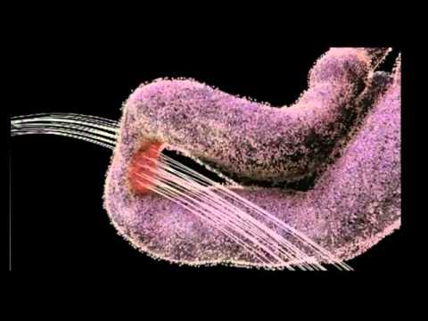
呼喵~ が 2021 年 01 月 14 日 に投稿私たちの細胞の中で、小さな分子マシンはどのように機能しているか、不思議に思ったことはありませんか?この動画では、DNA複製とマラリアのライフサイクルという驚くべき世界を、英語力アップに最適な高度な科学用語満載で解説します。驚異的な分子アニメーションを見ながら、たくさんの新しい単語を自然と身につけられるはずですよ!
この動画をアプリで学ぼう!
VoiceTubeアプリ版なら、効果的な学習機能がもっと充実しています!