字幕と単語
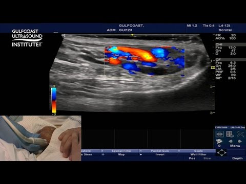
ホットなヒント - 下肢静脈瘤のための精巣超音波検査 (Hot Tips - Testicular Ultrasound for Varicocele)
00
jack が 2021 年 01 月 14 日 に投稿保存
動画の中の単語

to
US /tu,tə/
・
UK /tə/
- adv.に;に
- prep.(接触・結合・付着・付加を表して)~に(へ):~の上に:~に加えて;(比較を表して)~に比べて:~より;(方向を表して)~(のほう)へ;(行為・作用の対象を表して)~に対して:~に;(呼応を表して)~に答えて:~に応じて;…しに(行く);(限度・程度などを表して)~に至るまで:~するほどに;に;(感情を表す名詞を伴って)~したことには:~にも;に;(方向を表して)~に(へ):~まで;(到達点を表して)~まで:~に至るまで;(関係を表して)~の:~にとっての;~に向かって : ~のほうへ;(時間・期限の終わりを表して)~まで;(随伴を表して)~に合わせて:~について
- particle~する
A1 初級
もっと見る great
US /ɡret/
・
UK /ɡreɪt/
- adv.非常によく : すばらしく
- adj.大きい;重要な;すばらしい;素晴らしい;上手な;大
- n. (c.)偉大な : 卓越した
A1 初級TOEIC
もっと見る エネルギーを使用
すべての単語を解除
発音・解説・フィルター機能を解除

